


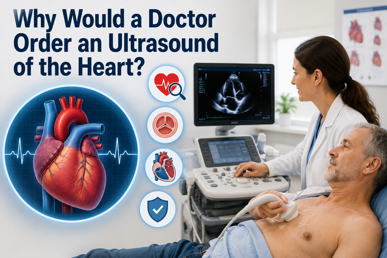
A doctor may order an ultrasound of the heart, medically known as an echocardiogram, to closely examine how well the heart is working. This painless imaging test uses sound waves to create a moving picture of the heart’s chambers, valves, and blood flow.
Physicians rely on this scan to detect problems that cannot always be found through a physical exam alone. It helps confirm suspected heart conditions, track existing ones, or rule out issues when a patient reports unusual symptoms.
Understanding why would a doctor order an ultrasound of the heart can help patients feel more prepared and less anxious before the appointment.
What Is a Heart Ultrasound?
A heart ultrasound, or echocardiogram, is a non-invasive scan that shows the heart’s structure and function in real time. It gives doctors a clear internal view without needles, radiation, or surgery involved.
How Echocardiogram Works?
The test uses a device called a transducer, which sends high-frequency sound waves toward the chest. These waves bounce off the heart’s tissues and return as echoes, which a computer then converts into a moving image. This allows the doctor to watch the heart beat, pump blood, and open its valves in real time.
Echocardiogram vs Other Heart Tests
Unlike an electrocardiogram (ECG), which only records the heart’s electrical activity, an echocardiogram shows its physical structure and motion. It also differs from a CT scan or angiogram since it involves no radiation or dye injection. This makes it a safer first-choice test for most heart-related concerns.
Types of Heart Ultrasound Tests
Not every echocardiogram is performed the same way. Doctors choose a specific type depending on what they need to examine and how detailed the images must be.
| Type of Echocardiogram | Description | Common Use |
| Transthoracic Echocardiogram (TTE) | Standard scan performed on the chest surface | General heart evaluation |
| Transesophageal Echocardiogram (TEE) | Probe passed through the esophagus for closer images | Detailed valve or clot assessment |
| Stress Echocardiogram | Performed during or after physical exertion | Checking blood flow under stress |
| Fetal Echocardiogram | Used on unborn babies during pregnancy | Detecting congenital heart defects |
| Doppler Echocardiogram | Measures speed and direction of blood flow | Identifying blockages or leaks |
Main Reasons Doctors Order This Test
There are many clinical situations where a heart ultrasound becomes necessary. Each reason relates to a specific concern about the heart’s structure, rhythm, or performance.


Checking Heart Pumping Strength
Doctors often use this test to measure the ejection fraction, which shows how much blood the heart pumps with each beat. A low ejection fraction can indicate weakened heart muscle or early heart failure. This measurement helps guide treatment decisions and future monitoring.
Evaluating Valve Problems
Heart valves can become narrow, stiff, or leaky over time, disrupting normal blood flow. An echocardiogram lets doctors see exactly how the valves open and close during each heartbeat. This helps determine whether medication, monitoring, or surgery is the right next step.
Detecting Structural Defects
Some people are born with structural heart defects that go unnoticed until symptoms appear later in life. This scan can reveal holes between chambers, unusual chamber sizes, or abnormal connections. Early detection allows for timely and appropriate treatment planning.
Investigating Symptoms
When a patient reports chest pain, breathlessness, dizziness, or fatigue, doctors often order this scan to find the underlying cause. It helps rule out or confirm heart-related reasons behind these symptoms. This is one of the most common reasons the test is requested in clinics.
After a Heart Attack
Following a heart attack, doctors need to know how much heart muscle was affected and how well it is recovering. An echocardiogram shows areas of damaged tissue and how they impact overall pumping ability. This information guides recovery plans and medication adjustments.
Monitoring Heart Failure
Patients already diagnosed with heart failure require regular monitoring to track disease progression. Repeated echocardiograms help doctors see whether the condition is stable, improving, or worsening. This ongoing tracking supports better long-term management.
Checking for Clots or Tumors
Occasionally, blood clots or unusual growths can form inside the heart chambers, increasing the risk of stroke or other complications. This scan can detect such masses with clear visual confirmation. Identifying them early allows doctors to begin appropriate treatment quickly.
Detecting Fluid Around Heart
Excess fluid buildup around the heart, known as pericardial effusion, can restrict normal heart movement. An echocardiogram clearly shows this fluid and measures its severity. This helps doctors decide if drainage or further intervention is required.
Evaluating Irregular Heartbeat
Patients experiencing arrhythmias, or irregular heart rhythms, often undergo this scan to check for underlying structural causes. It helps doctors understand whether the rhythm issue stems from valve disease, enlarged chambers, or other conditions. This insight shapes the treatment approach going forward.
Pre-Surgery Evaluation
Before major surgeries, doctors sometimes order this test to confirm the heart can safely handle the procedure and anesthesia. It provides a baseline understanding of heart function beforehand. This step reduces surgical risks and helps the medical team prepare accordingly.
Symptoms That May Lead to This Test
Certain warning signs commonly prompt doctors to recommend a heart ultrasound. These include:
- Persistent chest pain or tightness
- Shortness of breath during rest or activity
- Ongoing fatigue or unexplained weakness
- Swelling in the legs, ankles, or feet
- Noticeable heart palpitations or skipped beats
- Frequent dizziness or fainting spells
If any of these symptoms appear regularly, a doctor may suggest this scan to investigate further.
Who Typically Needs This Test?
While anyone with heart-related symptoms may need this scan, certain groups are more commonly referred for it. These include individuals with existing risk factors, family history of heart disease, or abnormal findings during routine checkups.
- People with high blood pressure or diabetes affecting heart health
- Athletes undergoing pre-participation heart screening
- Pregnant women needing fetal heart evaluation
- Patients with abnormal ECG readings
- Individuals with a heart murmur detected during examination
How the Test Is Performed?
The procedure is straightforward and generally takes less than an hour to complete in a clinical setting.
Step-by-Step Process
The patient lies on an exam table while a technician applies gel to the chest area. A handheld transducer is then moved across the skin to capture different angles of the heart. Images appear instantly on a monitor, allowing real-time observation throughout the scan.
Duration and Preparation
Most standard echocardiograms take between thirty and sixty minutes, depending on complexity. No special preparation is usually required, though a TEE may require fasting beforehand. Patients can typically resume normal activities immediately afterward.
Understanding the Results
After the scan, doctors review specific measurements to assess overall heart health. The table below outlines commonly evaluated results.
| Measurement | What It Shows |
| Ejection Fraction | Pumping efficiency of the heart |
| Chamber Size | Whether chambers are enlarged or normal |
| Valve Function | How well valves open and close |
| Wall Motion | Movement of heart muscle segments |
| Blood Flow Pattern | Direction and speed of blood through the heart |
These results help doctors decide whether further testing, medication, or lifestyle changes are necessary.
Is It Safe?
An echocardiogram is considered one of the safest heart tests available since it does not use radiation or invasive tools for the standard version. The transesophageal version may cause mild throat discomfort, and sedation carries minimal standard risks.
Overall, complications are extremely rare, making this test suitable for most patients, including children and pregnant women.
Frequently Asked Questions
How long does the test take? Most standard scans are completed within thirty to sixty minutes, though timing can vary based on individual cases.
Do I need to fast before the test? Fasting is generally not required for a standard scan but may be necessary before a transesophageal version.
Can I drive myself home afterward? Yes, in most cases, unless sedation was used during a transesophageal procedure.
How often should I get one if I have heart disease? This depends on your condition’s severity, and your cardiologist will recommend a suitable monitoring schedule.
Is this the same as an ECG? No, an ECG measures electrical activity, while this test shows the heart’s physical structure and motion.
Conclusion
Understanding why would a doctor order an ultrasound of the heart helps patients approach the test with more confidence and less worry.
From checking pumping strength to detecting valve issues, structural defects, or fluid buildup, this scan offers valuable insight into overall heart health. It remains a safe, painless, and highly informative tool that supports accurate diagnosis and effective treatment planning across nearly every stage of cardiac care.
Related Posts
Sudden Heel Pain Without Injury: Causes, Symptoms and Relief
What Do Kidney Stones Look Like in the Toilet? (Full Guide)
Can a Toothache Cause a Sore Throat? A Dentist Explains the Connection
Heart Home Health Care: Complete Guide to Services, Care Process, and Benefits